How the developing brain grows the protective insulation
The brain at birth
At birth, relatively few neuro-pathways have the fatty insulation coating called myelin. In brain development the insulation(1) process called myelination continues from before birth until about 20 years old. Until the age of 10 or so, vast areas of the cerebral cortex are not yet insulated with myelin. Up to the age of 20 large areas of the frontal lobes are not yet myelinated*.
Myelin is made up of a variety of insulating fats called lipids in a mixture which changes over time. The composition of myelin in an infant is quite different from that of an adult (and infants’ nerve impulses travel more slowly). The supply of lipids for myelin and the changes in myelin composition as the brain matures are provided by lipid processing enzymes. These enzymes are essential for the maintenance of normal myelin on all the nerve axons.
The assembly of myelin sheaths is the highly specialized function of oligodendrocytes in the central nervous system and Schwann cells in the peripheral nervous system. Myelin-forming glial cells wrap axons with multiple layers of fatty membrane and provide the electrical insulation that is necessary for a “rapid impulse” propagation.
White matter gray matter
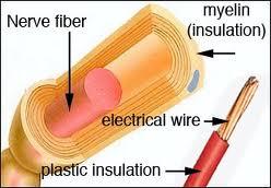
The white matter is the areas of the brain and nervous system rich in axons and dendrites. Axons are like electrical wires along which neural messages travel away from the cells and dendrites carry messages towards the cell. These messages translate to muscle movement, hearing, sight, etc. In the brain maturation process the axons and dendrites are covered with glial cells. The glial cells are supportive cells like the colored plastic insulation we see around electrical wires. The messages (which are similar to an electric current) travel down the axons and up the dendrites. The myelin helps keep the signal from becoming mixed up in the other message pathways.
Different parts of the nervous system appear gray, white, or mottled. The gray matter is made up of a mix of cells and capillary blood vessels and has a gray appearance. As we already stated, the connections between the cells are the axons covered in glial cells. The glial cells, being fatty, have a high refractive index and appear white. An area of the brain rich in these is called the white matter. A part of the brain which is a good mix of cell bodies and axons (gray and white matter) is called the reticular formation as it has a netlike appearance.
Brain and nerve conduction
The parent cell body which hosts the axons and dendrites is called the oligodendrocyte. A single oligodendrocyte may be responsible for up to 20 myelin segments which are organized like the tentacles of an octopus. The wrap is like a Swiss roll, with as many of 30 turns of sheath. The sheaths butt to the nodes of Ranvier. Conduction is along the sheath and is known as “saltatory.” A non myelinated fibre may conduct at about 0.5 meters a second where a fully myelinated fiber of the same size may conduct at 150 meters a second.
A further insulation in the nervous system is provided by cerebrospinal fluid(CSF). The CSF is rich in surfactant which absorbs to the surface of the myelin sheath. The CFS is a Zwitterion, or a strongly charged ion that is negative at one end and positive at the other thereby sending the signals back to the myelin.
Fibers in gray matter may also be myellinated. The oligodendrocyte has a very high metabolic rate and the sheaths are easily damaged by edema where there is protein extravasation (due to complement activation which is a complex system of proteins designed to eliminate infectious microorganisms) or they are damaged by a chemical or metallic insult.
1.Even though we liken the way the myelin sheath works to insulation, it works by increasing the speed of message transmission by saltatory conduction. The impulses pass along the outer aspect of the sheath longitudenally and across the junctions known as the nodes of Ranvier.
- [Ref: Peter Nathan, The Nervous System, (Philadelphia: J.B. Lippincott, 1969), p.296.;
- Leslie Hart, Human Brain and Human Learning, (New York: Longman Inc., White Plains) Books for Educators, Oak Creek, CA),p.119]
Read more in-depth with this book series: Fixing The Brain by Craig Stellpflug
Authored by Neurodevelopment Consultant Craig Stellpflug NDC, CNC, Healing Pathways Medical Clinic Scottsdale, AZ
Copyright 2012 Craig Stellpflug© Permission is hereby granted to copy and distribute this article but only in its entirety